Gut-Derived Triggers of Multiple Sclerosis

MultipleSclerosis GutMicrobiome AutoimmuneDisease Immunology KeioUniversity Neuroinflammation

Riepilogo

Immagine Copertina Mappa Mentale
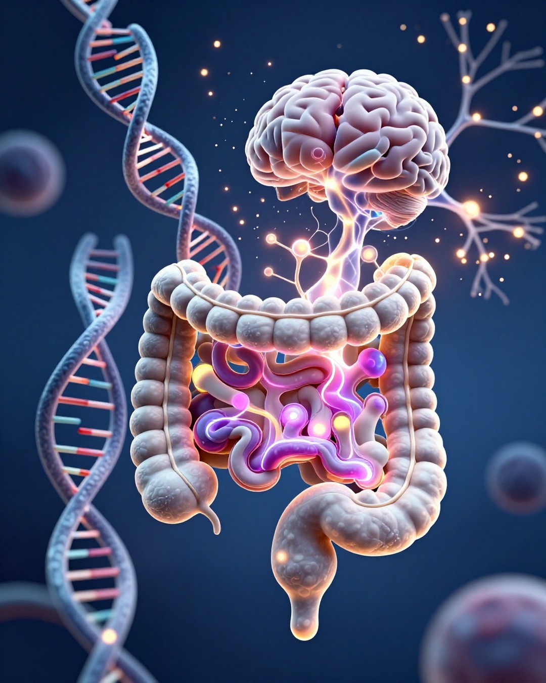
Immagine Copertina Generata da AI

Researchers from Keio University, led by Dr. Shohei Suzuki and Dr. Tomohisa Sujino, have identified a critical gut-CNS axis that triggers multiple sclerosis (MS). Published on June 25, 2026, in Science Immunology, the study reveals that intestinal epithelial cells (IECs) play an active role in autoimmune inflammation by presenting antigens to CD4+ T cells, which then polarize into pathogenic Th17 cells and migrate to the central nervous system. The authors demonstrate that this mechanism is conserved across both experimental autoimmune encephalomyelitis (EAE) mouse models and human patients. By showing that MHC II expression in the gut is essential for the generation of these neuroinflammatory cells, the research shifts the understanding of MS from a purely neurological condition to one with significant gastrointestinal origins. This discovery provides a clear cellular mechanism: IECs upregulate MHC II in response to signals, directly priming T cells in the gut mucosa. These activated Th17 cells subsequently travel through the systemic circulation to the brain and spinal cord, where they attack myelin sheaths, causing the characteristic damage associated with MS. The findings offer promising therapeutic avenues, suggesting that modulating the gut microbiome or inhibiting the antigen-presenting activity of intestinal epithelial cells could serve as novel strategies for treating autoimmune neurological diseases. This research underscores the importance of the gut-brain connection in managing complex autoimmune disorders.

Punti Chiave

01

🔹 The Gut-CNS Axis Mechanism

【📌 Core Finding】Intestinal epithelial cells (IECs) act as active participants in MS by presenting antigens to CD4+ T cells. 【🔸 Process】IECs upregulate MHC II (Major Histocompatibility Complex Class II) molecules, which prime CD4+ T cells and polarize them into pathogenic Th17 cells. These cells then migrate from the intestinal lamina propria to the central nervous system, driving neuroinflammation.

02

🔸 Evidence from EAE and Human Patients

【⭐ Data Support】Researchers utilized single-cell RNA sequencing on intestinal biopsies from MS patients and EAE mouse models. Both groups exhibited a significant accumulation of inflammatory Th17 cells in the small intestine. The study confirmed that deleting MHC II in IECs significantly reduced the generation of pathogenic Th17 cells and decreased overall disease severity.

03

⭐ Role of MHC II in Pathogenesis

【📌 Key Finding】Under normal conditions, IECs do not typically present antigens. However, in the context of MS, they become active antigen-presenting cells. Co-culture assays demonstrated that these cells directly interact with CD4+ T cells, pushing them toward a pro-inflammatory Th17 phenotype, which is a hallmark of the autoimmune response in MS.

04

📊 Tracking Cell Migration

【📈 Methodology】To verify the migration of these cells, the team used transgenic mice expressing the Kaede protein. This allowed for the visual tracking of pathogenic Th17 cells from the intestinal lamina propria to the spinal cord. The results provided definitive evidence that gut-derived immune cells directly contribute to the autoreactive cell population in the CNS.

05

💡 Therapeutic Implications

【🎯 Future Outlook】Current MS therapies primarily target B cells. This study suggests that the gut is a viable therapeutic site. Potential treatments could involve modulating the gut microbiota or specifically inhibiting the antigen-presenting activity of intestinal epithelial cells to prevent the formation of encephalitogenic T cells before they reach the brain.

Vuoi creare la tua mappa mentale?

Accedi a Neumap AI e inizia a creare le tue mappe mentali!

Inizia Ora