DNA Folding Visualization Breakthrough

scitechdaily biology genetics dna-folding microscopy

Resumo

Imagem de Capa do Mapa Mental
Imagem de Capa Gerada por IA

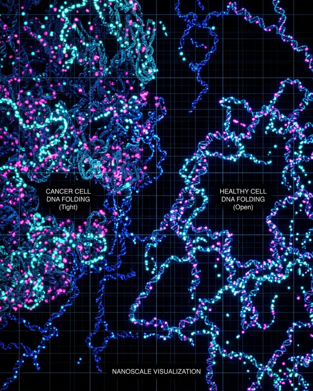
Scientists have developed innovative fluorescent probes called HoTs that enable the visualization of DNA folding and movement inside living cells at exceptionally fine scales. Published on September 3, 2026, the study from the Centre for Genomic Regulation shows that these auto-blinking dyes can pinpoint individual molecules to within 3 nanometers in preserved cells and 20 nanometers in living cells using advanced microscopy techniques like MINFLUX and STORM. The research highlights a striking difference in cancer cells from human bowel tissue, where DNA appears noticeably looser and more spread out compared to healthy tissue. This progressive unpacking as cancer develops suggests that the degree of DNA folding could eventually serve as a critical diagnostic biomarker for determining tumor aggressiveness and staging. The breakthrough overcomes traditional limitations where powerful microscopy required harsh chemicals and dead cells, enabling real-time observation of chromatin behavior. By combining the HoTs probes with the AI program AINU, researchers successfully distinguished skin cells from stem cells with 96 to 98 percent accuracy based on nuclear DNA packaging patterns. This technology bridges a major gap in structural biology by offering non-invasive insights into how packed DNA controls gene activation. Future applications could revolutionize disease diagnostics, cancer prognostics, and the identification of optimal stem cells for regenerative medicine therapies.

Pontos-chave

01

🔹 HoT Auto-Blinking Fluorescent Probes

【📌 Core Innovation】Researchers developed HoTs, novel fluorescent dyes that enter living cells independently and bind to DNA. 【⭐ Mechanism】Unlike traditional fluorophores that glow continuously, HoTs blink intermittently, switching on and off to prevent signal blurring. 【📊 Precision Metrics】Using STORM microscopy, the probes visualize living cell DNA at 20 nanometer resolution, while MINFLUX achieves 3 nanometer precision in preserved cells, nearing the 2 nanometer width of the DNA double helix.

02

🔸 Cancer Cell DNA Unpacking Discovery

【📌 Diagnostic Finding】Testing wax-preserved bowel tissue from three cancer patients revealed distinct structural differences in nuclear organization. 【⭐ Structural Contrast】DNA in tumor cells is noticeably looser and more spread out compared to tight, distinct packages found in healthy adjacent tissue. 【📈 Clinical Implication】The progressive unpacking of DNA during cancer development suggests folding degrees could serve as future markers for tumor aggressiveness.

03

⭐ Integration with AINU Artificial Intelligence

【📌 AI Analysis】The team utilized AINU (AI of the Nucleus), a program capable of detecting subtle patterns in super-resolution nuclear images. 【📊 Accuracy Data】After training on new living cell images, AINU distinguished skin cells from stem cells with 96 to 98 percent accuracy. 【🔗 Functional Link】Despite containing identical DNA, different cell types package chromatin uniquely, allowing the AI to successfully classify them.

04

📊 Cross-Species and Tissue Applications

【📌 Zebrafish Study】Probes were applied to zebrafish eye tissue to study retinal regeneration processes where cells loosen DNA to gain flexibility. 【⭐ Broad Utility】The methodology successfully bridges observations across human skin cells, HeLa cancer cell lines, and vertebrate model tissues. 【💡 Research Lead】Senior author Pia Cosma and co-senior author Hongyan Sun published these findings on August 28, 2026, in the journal Molecular Cell.

05

💡 Technical Limitations and Future Directions

【📌 Current Constraints】The fluorescent molecules coat DNA broadly rather than targeting specific individual genes, and highest-resolution images still require preserved cells. 【🔄 Future Objectives】Researchers are currently working to optimize MINFLUX to achieve maximum resolution in entirely live cells. 【🛡️ Medical Impact】Combining HoTs and AI aims to eventually automate cancer versus healthy tissue identification and advance regenerative medicine.

Quer criar seu próprio mapa mental?

Faça login no Neumap AI e comece a criar seus próprios mapas mentais!

Começar