Muscle Cells in Bone Fracture Repair

BoneResearch BoneHealing StemCells OrthopedicResearch CellBiology

Summary

Mind Map Cover Image
AI Generated Cover Image

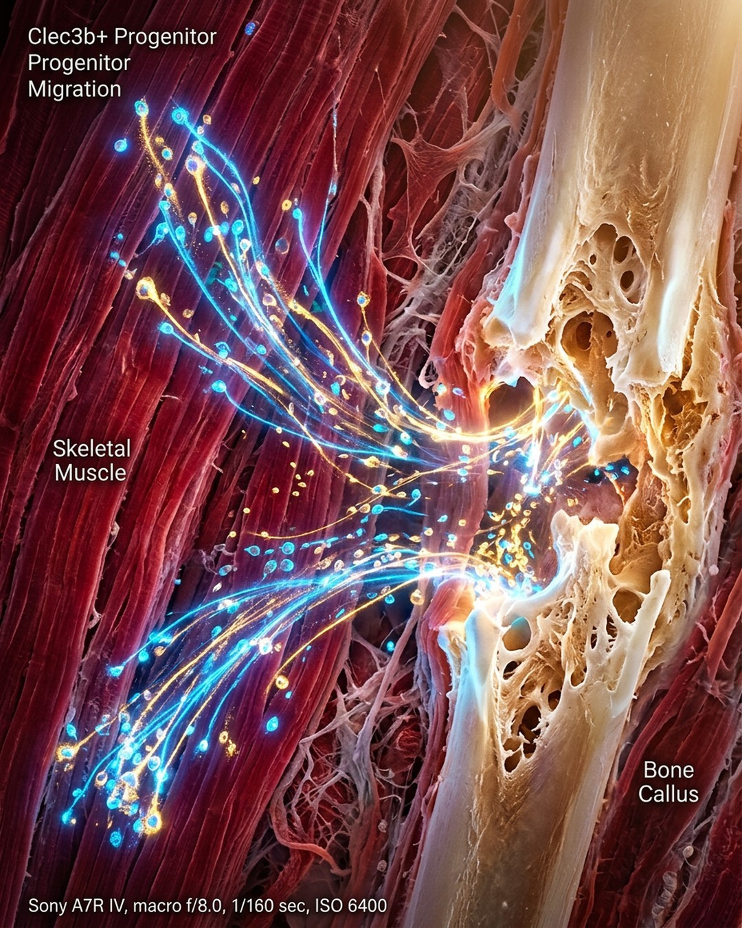
A study published in Volume 14 of the journal Bone Research on July 06, 2026, led by Dr. Ugur M. Ayturk and colleagues from the Skeletal Health and Orthopedic Research Program in the USA, revealed that muscle-resident fibroadipogenic progenitors (FAPs) and a smaller population of superficial periosteal cells play an unexpected and vital role in repairing fractured bones. The researchers engineered a mouse model utilizing Clec3b expression as a highly specific marker to track these dormant progenitor cells under normal and injured conditions, discovering that they rapidly migrate to fracture sites and differentiate into osteoblasts and bone marrow stromal cells. Within three weeks following a fracture, approximately 28% of the osteoblasts in the healing callus originated from Clec3b-lineage cells, with skeletal muscle identified as the primary source of these regenerative cells compared to the periosteum. These cells also contribute to heterotopic ossification, where bone abnormally forms in soft tissues, suggesting that therapeutically activating or limiting Clec3b-lineage cells could enhance fracture healing or prevent unwanted abnormal bone growth after injuries.

Key Points

01

🔹 Discovery of Extraskeletal Progenitor Cells in Bone Repair

【📌 Key Finding】Published on July 06, 2026, in Volume 14 of the journal Bone Research, researchers led by Dr. Ugur M. Ayturk identified that muscle-resident fibroadipogenic progenitors (FAPs) and superficial periosteal cells remain dormant under normal conditions but acquire bone-forming functions after injury. 【⭐ Data Support】Clec3b expression served as a highly specific marker to track these progenitor cells using an engineered mouse model with a fluorescent tag.

02

🔸 Migration and Differentiation Mechanisms Post-Injury

【📌 Process Steps】Dormant Clec3b-lineage cells migrated rapidly to fracture sites following bone injury, where they differentiated into osteoblasts and bone marrow stromal cells. 【⭐ Data Support】Within three weeks, approximately 28% of the osteoblasts in the healing callus originated from Clec3b-lineage cells. The cells stopped expressing Clec3b as they differentiated, confirming their transition from a dormant progenitor state.

03

⭐ Skeletal Muscle as the Primary Source of Regenerative Cells

【📌 Comparative Analysis】Experiments comparing skeletal muscle and the periosteum demonstrated that skeletal muscle is the main source of these bone-forming cells. 【⚖️ Key Differences】Even after surgical removal of the periosteum, Clec3b-positive cells reached the fracture site. Furthermore, bone grafts including surrounding muscle generated substantially more Clec3b-lineage bone-forming cells than grafts without muscle.

04

📊 Contribution to Heterotopic Ossification and Pathological Bone Growth

【📌 Core Finding】Clec3b-lineage cells also contribute to heterotopic ossification, a condition where bone abnormally forms in muscles and soft tissues following injury. 【🔗 Cause & Effect】Blocking key pathways required for bone formation or depleting these cells significantly reduced both fracture healing and abnormal bone growth, proving their dual role in normal and pathological bone remodeling.

05

💡 Therapeutic Potential and Future Applications

【📌 Application Value】Dr. Ugur M. Ayturk and colleagues noted that these cells represent promising therapeutic targets for clinical treatments. 【🛡️ Prevention & Enhancement】Activating these progenitor cells could strategically enhance fracture healing in patients, while limiting their bone-forming activity could prevent unwanted heterotopic ossification following serious physical injuries.

Want to create your own mindmap?

Login to Neumap AI and start creating your own mindmaps!

Get Started